Therapeutic Targets and Technological Innovations Axis

Field of research
This axis focuses on innovations related to the treatment of pain in patients, and technological innovations developed in the form of tools for preclinical research.
Transcranial magnetic stimulation (rTMS) of the primary motor cortex at high frequency (>5 Hz) induces analgesic effects, probably by activating pain modulation systems. We show that a new rTMS paradigm, using continuous theta stimulation (pcTBS), composed of bursts of three pulses at 50 Hz repeated five times per second
for 1 min and 44 s (1,200 pulses), is significantly more effective than rTMS at 10 Hz (Moisset et al. Brain Stim 2015). The pcTBS protocol could therefore have considerable clinical potential, being 8 times shorter but producing stronger analgesic effects than classic rTMS.
Source : Moisset et al. Eur J Neurol. 2017
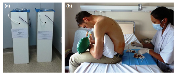
Lumbar puncture (LP) has been frequently performed for over a century. This procedure is always stressful and often painful. A randomized controlled trial was conducted in adults who required cerebrospinal fluid analysis to evaluate the effectiveness of a fixed mixture of nitrous oxide and 50% oxygen compared to placebo to reduce immediate pain and anxiety procedures during LP (Moisset et al. Eur J Neurol. 2017). We found that inhalation of a fixed 50% N2 O-O2 mixture is effective in reducing PL-induced pain and anxiety. In addition to innovations for patients, several tools have been developed by the laboratory team, with the aim of automating certain repetitive and time-consuming tasks, and therefore reducing inter and intra operator variations. The major needs mainly concern video ethology techniques, or calcium imaging or confocal imaging. In the case of imaging, this involves detecting and counting the neurons in a slice or section of the brain or spinal cord. These neurons are visible either because they have been marked with a fluorescent antibody or because they have been genetically modified to fluoresce upon absorption of calcium.
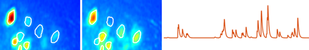
Delineation of neurons in calcium imaging and deconvolved neuronal activity signal
In the case of calcium imaging, the tool transforms a dynamic 3D image (2D+time) into temporal neuronal activity. The deconvolution operation uses the FOOPSI approach, a constrained sparse deconvolution.
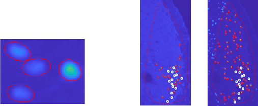
Neural detection by shape optimization
In the case of confocal imaging and fluorescent labeling, detection uses a parametric ellipsoidal model and unconstrained minimization, with an approach resolutely based on the shape of neurons, rather than on fluorescence intensity.
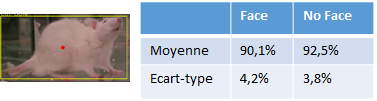
Finally, with the aim of automating the application of the grimace scale in the rating of spontaneous painful behaviors, the team is investing in the disciplinary field of supervised learning, initially to automatically detect the presence of the face of an animal. Preliminary results show excellent detection rates (>90%).
Faculty
-
 Cristina ALBA DELGADO, Assistant Professor, Team TPM – Pathophysiology of migraine axis, Team Trigeminal Pain and MigraineCristina.ALBA_DELGADO@-Code to remove to avoid SPAM-uca.fr, +33 4 73 17 73 20, room 246
 -
 Myriam ANTRI, Assistant Professor, Team TPM – Pathophysiology of mechanical allodynia axis, Team TPM – Pathophysiology of migraine axis, Team TPM – Therapeutic Targets and Technological Innovations Axis, Team Trigeminal Pain and Migrainemyriam.antri@-Code to remove to avoid SPAM-uca.fr, +33 4 73 17 73 14, room 241
 -
 Denis ARDID, Professor, Fundamental Pharmacology and Pain Clinic Team, Team FPPC – Microbial Metabolites and Chronic Pain Axisdenis.ardid@-Code to remove to avoid SPAM-uca.fr, +33 4 73 17 82 39
 -
 Alain ARTOLA, Professor Emeritus HDR, Team TPM – Pathophysiology of mechanical allodynia axis, Team Trigeminal Pain and MigraineAlain.ARTOLA@-Code to remove to avoid SPAM-uca.fr, +33(0)4-7317-7312
 -
 Nicolas AUTHIER, Professor, Fundamental Pharmacology and Pain Clinic Team, Team FPPC- Axe PatientNicolas.AUTHIER@-Code to remove to avoid SPAM-uca.fr
 -
 David BALAYSSAC, Professor, Fundamental Pharmacology and Pain Clinic Team, Team FPPC – Pain and Cancer Axisdavid.balayssac@-Code to remove to avoid SPAM-uca.fr, +33 4 73 17 80 41
 -
 Isabelle BARTHELEMY, Professor, Team Trigeminal Pain and Migraineibarthelemy@-Code to remove to avoid SPAM-chu-clermontferrand.fr
 -
 Mélina BEGOU, Assistant Professor, Fundamental Pharmacology and Pain Clinic Team, TEAM 2 – Molecular and Cellular Pain Modulators AxisMelina.BEGOU@-Code to remove to avoid SPAM-uca.fr, + 33 4 73 17 81 02
 -
 Célian BERTIN, Medical Assistant, Fundamental Pharmacology and Pain Clinic Team, Team FPPC- Axe Patientcelian.bertin@-Code to remove to avoid SPAM-uca.fr
 -
 Jérôme BUSSEROLLES, Assistant Professor, Fundamental Pharmacology and Pain Clinic Team, Team FPPC – Pain and Cancer AxisJerome.BUSSEROLLES@-Code to remove to avoid SPAM-uca.fr, +33(0)4-7317-8235
 -
 Frédéric CARVALHO, Research Scientist, Fundamental Pharmacology and Pain Clinic Team, Team FPPC – Microbial Metabolites and Chronic Pain Axisfrederic.carvalho@-Code to remove to avoid SPAM-uca.fr
 -
 Chouki CHENAF, Assistant Professor, Fundamental Pharmacology and Pain Clinic Team, Team FPPC- Axe Patientchouki.chenaf@-Code to remove to avoid SPAM-uca.fr
 -
 David CIA, Assistant Professor, Fundamental Pharmacology and Pain Clinic Team, Team FPPC – Microbial Metabolites and Chronic Pain Axisdavid.cia@-Code to remove to avoid SPAM-uca.fr, +33 (0)4 73 17 82 30 (33) 4 73 17 79 83
 -
 Pierre CLAVELOU, Professor HDR, Team TPM – Pathophysiology of migraine axis, Team Trigeminal Pain and MigrainePierre.CLAVELOU@-Code to remove to avoid SPAM-uca.fr
 -
 Aurore COLLIN, Assistant Professor, Fundamental Pharmacology and Pain Clinic Team, Team FPPC – Pain and Cancer Axisaurore.collin@-Code to remove to avoid SPAM-uca.fr
 -
 Christine COURTEIX, Professor, Fundamental Pharmacology and Pain Clinic Team, TEAM 2 – Molecular and Cellular Pain Modulators Axischristine.courteix@-Code to remove to avoid SPAM-uca.fr
 -
Cristelle Dalbos, Professor Emeritus, Fundamental Pharmacology and Pain Clinic Team
-
 Radhouane DALLEL, Professor HDR, Team TPM – Pathophysiology of mechanical allodynia axis, Team TPM – Pathophysiology of migraine axis, Team TPM – Therapeutic Targets and Technological Innovations Axis, Team Trigeminal Pain and MigraineRadhouane.DALLEL@-Code to remove to avoid SPAM-uca.fr, +33(0)4-7317-7313, room 239
 -
-
Noémie Delage, Professor, Fundamental Pharmacology and Pain Clinic Team, Team FPPC- Axe Patientndelage@-Code to remove to avoid SPAM-chu-clermontferrand.fr
-
 Lauriane DELAY, Assistant Professor, Fundamental Pharmacology and Pain Clinic Team, TEAM 2 – Molecular and Cellular Pain Modulators Axislauriane.delay@-Code to remove to avoid SPAM-uca.fr
 -
 Christophe DESCHAUMES, Assistant Professor, Team TPM – Pathophysiology of mechanical allodynia axis, Team Trigeminal Pain and MigraineChristophe.DESCHAUMES@-Code to remove to avoid SPAM-uca.fr, +33(0)4-7317-7316, room 240
 -
 Laurent DEVOIZE, Professor HDR, Team TPM – Pathophysiology of mechanical allodynia axis, Team Trigeminal Pain and MigraineLaurent.DEVOIZE@-Code to remove to avoid SPAM-uca.fr, +33(0)4-7317-7316, room 240
 -
 Stéphane Doly, Assistant Professor, Fundamental Pharmacology and Pain Clinic Team, TEAM 2 – Molecular and Cellular Pain Modulators Axisstephane.doly@-Code to remove to avoid SPAM-uca.fr
 -
 Anne DONNET, Medical Doctor, Team TPM – Pathophysiology of migraine axis, Team TPM – Therapeutic Targets and Technological Innovations Axis, Team TPM -Technical Advances, Team Trigeminal Pain and MigraineAnne.DONNET@-Code to remove to avoid SPAM-ap-hm.fr
 -
 Christian DUALE, Medical Doctor, Team TPM – Pathophysiology of mechanical allodynia axis, Team Trigeminal Pain and Migrainecduale@-Code to remove to avoid SPAM-chu-clermontferrand.fr
 -
 Claude Dubray, Professor Emeritus, Fundamental Pharmacology and Pain Clinic Team, Team FPPC- Axe PatientClaude.DUBRAY@-Code to remove to avoid SPAM-uca.fr
 -
 Anne DUTOUR, Medical Doctor, Team TPM – Pathophysiology of mechanical allodynia axis, Team TPM – Therapeutic Targets and Technological Innovations Axis, Team Trigeminal Pain and Migraineanne.dutour@-Code to remove to avoid SPAM-uca.fr, +33 4 73 17 73 12, room 252
 -
 Alain Eschalier, Professor Emeritus, Fundamental Pharmacology and Pain Clinic Teamalain.eschalier@-Code to remove to avoid SPAM-uca.fr
 -
 Camille FAUCHON, Professor, Team TPM – Pathophysiology of migraine axis, Team TPM – Therapeutic Targets and Technological Innovations Axis, Team Trigeminal Pain and Migrainecamille.fauchon@-Code to remove to avoid SPAM-uca.fr, +33 6 99 24 57 90, room 249
 -
Florent Ferrer, Medical Doctor, Fundamental Pharmacology and Pain Clinic Team, Team FPPC- Axe Patientfferrer@-Code to remove to avoid SPAM-chu-clermontferrand.fr
-
 Agathe GELOT, Assistant Professor, Fundamental Pharmacology and Pain Clinic Team, Team FPPC – Microbial Metabolites and Chronic Pain Axisagathe.gelot@-Code to remove to avoid SPAM-uca.fr
 -
 Fabrice Giraudet, Assistant Professor, Fundamental Pharmacology and Pain Clinic Team, Team FPPC – Pain and Cancer Axisfabrice.giraudet@-Code to remove to avoid SPAM-uca.fr
 -
 Christelle GREMEAU-RICHARD, Assistant Professor, Team TPM – Pathophysiology of mechanical allodynia axis, Team TPM – Therapeutic Targets and Technological Innovations Axis, Team TPM -Technical Advances, Team Trigeminal Pain and Migrainechristelle.gremeau-richard@-Code to remove to avoid SPAM-uca.fr, +33 4 73 17 73 18, room 242
 -
Virginie Guastella, Medical Doctor, Fundamental Pharmacology and Pain Clinic Team, Team FPPC- Axe Patientvguastella@-Code to remove to avoid SPAM-chu-clermontferrand.fr
-
 Nathalie GUY, Medical Doctor, Team TPM – Pathophysiology of migraine axis, Team Trigeminal Pain and Migrainenguy@-Code to remove to avoid SPAM-chu-clermontferrand.fr
 -
 Michel LANTERI-MINET, Medical Doctor, Team TPM – Pathophysiology of migraine axis, Team TPM – Therapeutic Targets and Technological Innovations Axis, Team TPM -Technical Advances, Team Trigeminal Pain and Migrainelanteri-minet.m@-Code to remove to avoid SPAM-chu-nice.fr
 -
 Stéphane LOLIGNIER, Assistant Professor, Fundamental Pharmacology and Pain Clinic Team, TEAM 2 – Molecular and Cellular Pain Modulators AxisStephane.LOLIGNIER@-Code to remove to avoid SPAM-uca.fr, +33 4 73 17 82 35
 -
 Philippe LUCCARINI, Professor HDR, Team TPM – Pathophysiology of migraine axis, Team TPM – Therapeutic Targets and Technological Innovations Axis, Team TPM -Technical Advances, Team Trigeminal Pain and Migrainephilippe.luccarini@-Code to remove to avoid SPAM-uca.fr, +33 4 7317 73 17, room 243
 -
 Christophe MALLET, Assistant Professor, Fundamental Pharmacology and Pain Clinic Team, Team FPPC – Pain and Cancer Axischristophe.mallet@-Code to remove to avoid SPAM-uca.fr
 -
 Fabien MARCHAND, Assistant Professor, Fundamental Pharmacology and Pain Clinic Team, TEAM 2 – Molecular and Cellular Pain Modulators Axisfabien.marchand@-Code to remove to avoid SPAM-uca.fr, +33(0)4-7317-8231
 -
 Sylvain MATHIEU, Medical Doctor, Team TPM – Pathophysiology of mechanical allodynia axis, Team Trigeminal Pain and Migrainesmathieu@-Code to remove to avoid SPAM-chu-clermontferrand.fr
 -
 Céline MELIN, Assistant Professor, Team TPM – Pathophysiology of mechanical allodynia axis, Team TPM – Therapeutic Targets and Technological Innovations Axis, Team TPM -Technical Advances, Team Trigeminal Pain and Migraineceline.melin@-Code to remove to avoid SPAM-uca.fr, +33 4 73 17 73 18, room 242
 -
 Xavier MOISSET, Professor HDR, Team TPM – Pathophysiology of mechanical allodynia axis, Team TPM – Pathophysiology of migraine axis, Team TPM – Therapeutic Targets and Technological Innovations Axis, Team TPM -Technical Advances, Team Trigeminal Pain and Migrainexavier.moisset@-Code to remove to avoid SPAM-uca.fr, +33 4 73 75 22 01
 -
 Thierry MOM, Professor, Team TPM – Pathophysiology of migraine axis, Team Trigeminal Pain and MigraineThierry.MOM@-Code to remove to avoid SPAM-uca.fr
 -
 Lénaic MONCONDUIT, Assistant Professor, Team TPM – Pathophysiology of migraine axis, Team TPM – Therapeutic Targets and Technological Innovations Axis, Team TPM -Technical Advances, Team Trigeminal Pain and Migrainelenaic.monconduit@-Code to remove to avoid SPAM-uca.fr, +33 4 73 17 73 14, room 241
 -
 Cedric PEIRS, Principal Investigator, Team TPM – Pathophysiology of mechanical allodynia axis, Team Trigeminal Pain and Migrainecedric.peirs@-Code to remove to avoid SPAM-inserm.fr, +33(0)4-7317-7311, room 245
 -
 Nathalie PHAM-DANG, Professor HDR, Team TPM – Pathophysiology of mechanical allodynia axis, Team Trigeminal Pain and Migrainenphamdang@-Code to remove to avoid SPAM-chu-clermontferrand.fr, +33 4 73 75 01 02, room 245
 -
 Gisèle PICKERING, Professor, Fundamental Pharmacology and Pain Clinic Team, Team FPPC – Pain and Cancer Axis, Team FPPC- Axe Patientgisele.pickering@-Code to remove to avoid SPAM-uca.fr
 -
 Isabelle RANCHON-COLE, Professor HDR, Team TPM – Pathophysiology of migraine axis, Team Trigeminal Pain and MigraineIsabelle.RANCHON-COLE@-Code to remove to avoid SPAM-uca.fr, +33(0)4-7317-1325, room 246
 -
Damien RICHARD, Medical Doctor, Fundamental Pharmacology and Pain Clinic Team, Team FPPC- Axe Patientdamien.richard@-Code to remove to avoid SPAM-uca.fr
-
Catherine Rubat-Coudert, Assistant Professor, Fundamental Pharmacology and Pain Clinic TeamCatherine.RUBAT-COUDERT@-Code to remove to avoid SPAM-uca.fr
-
 Laurent SAKKA, Assistant Professor, Fundamental Pharmacology and Pain Clinic Team, Team FPPC- Axe Patient
 -
-
 Benoît Sion, Assistant Professor, Fundamental Pharmacology and Pain Clinic Team, Team FPPC – Microbial Metabolites and Chronic Pain Axisbenoit.sion@-Code to remove to avoid SPAM-uca.fr
 -
 Eric WERSINGER, Assistant Professor, Fundamental Pharmacology and Pain Clinic Team, Team FPPC – Pain and Cancer Axiseric.wersinger@-Code to remove to avoid SPAM-uca.fr
 -
 Mickael ZBILI, Assistant Professor, Team TPM – Pathophysiology of mechanical allodynia axis, Team Trigeminal Pain and Migrainemickael.zbili@-Code to remove to avoid SPAM-uca.fr, +33 4 73 17 73 17, room 245
Support staff
-
 Youssef AISSOUNI, Senior Engineer, Fundamental Pharmacology and Pain Clinic Teamyoussef.aissouni@-Code to remove to avoid SPAM-inserm.fr, +33(0)4-7317-8231
 -
 Julie BARBIER, Assistant Engineer, Fundamental Pharmacology and Pain Clinic TeamJulie.BARBIER@-Code to remove to avoid SPAM-uca.fr
 -
 Clément BECQUIE, Assistant Engineer, Fundamental Pharmacology and Pain Clinic Team
 -
Christine CERCY, Technician, Fundamental Pharmacology and Pain Clinic Teamchristine.cercy@-Code to remove to avoid SPAM-uca.fr
-
Eric CHAPUY, Technician, Fundamental Pharmacology and Pain Clinic Teameric.chapuy@-Code to remove to avoid SPAM-uca.fr
-
Laurence CHAUVET, Technical assistant, Fundamental Pharmacology and Pain Clinic TeamLaurence.CHAUVET@-Code to remove to avoid SPAM-uca.fr
-
Emilie CLEMENCON, Administrative Assistant, Fundamental Pharmacology and Pain Clinic Teamemilie.clemencon@-Code to remove to avoid SPAM-uca.fr, +33 4 73 17 82 30
-
 Giséla DA SILVA BORGES, Engineer, Team Trigeminal Pain and Migrainegisela.da_silva_borges@-Code to remove to avoid SPAM-uca.fr, +33 4 73 17 73 67, room 244
 -
 Amélie DESCHEEMAEKER, Technician, Team Trigeminal Pain and Migraineamelie.descheemaeker@-Code to remove to avoid SPAM-uca.fr, +33 4 73 17 73 12, room 237
 -
 François GABRIELLI, Senior Engineer, Team TPM – Therapeutic Targets and Technological Innovations Axis, Team TPM -Technical Advances, Team Trigeminal Pain and Migrainefrancois.gabrielli@-Code to remove to avoid SPAM-uca.fr, +33 4 73 17 73 25, room 236
 -
Delphine Gauthier, Assistant Engineer, Fundamental Pharmacology and Pain Clinic TeamDelphine.GAUTHIER@-Code to remove to avoid SPAM-uca.fr
-
 Anne-Marie GAYDIER, Lab Manager, Team Trigeminal Pain and Migrainea-marie.gaydier@-Code to remove to avoid SPAM-uca.fr, +33 4 73 17 73 12, room 237
 -
 Anouk Giraudet, Assistant Engineer, Fundamental Pharmacology and Pain Clinic Team, Team FPPC – Microbial Metabolites and Chronic Pain Axisanouk.giraudet@-Code to remove to avoid SPAM-uca.fr
 -
 Karine HERAULT, Engineer, Team Trigeminal Pain and Migrainekarine.herault@-Code to remove to avoid SPAM-inserm.fr, +33 4 73 17 73 67, room 244
 -
Nathalie JACQUEMOT, Assistant Engineer, Fundamental Pharmacology and Pain Clinic Teamnathalie.jacquemot@-Code to remove to avoid SPAM-uca.fr
-
 Nicolas KERCKHOVE, Clinical Project Panager, Fundamental Pharmacology and Pain Clinic Team, Team FPPC- Axe Patientnicolas.kerckhove@-Code to remove to avoid SPAM-uca.fr
 -
Mathieu Meleine, Senior Engineer, Fundamental Pharmacology and Pain Clinic Teammathieu.meleine@-Code to remove to avoid SPAM-uca.fr
-
 Mireille MONTBEL, Technician, Fundamental Pharmacology and Pain Clinic Teammireille.montbel@-Code to remove to avoid SPAM-uca.fr
 -
 Victor Nowak, Assistant Engineer, Fundamental Pharmacology and Pain Clinic Team
 -
 Laetitia PRIVAL, Assistant Engineer, Fundamental Pharmacology and Pain Clinic Teamlaetitia.prival@-Code to remove to avoid SPAM-uca.fr
 -
 Sylviane ROUSSELIN, Caretaker, Team Trigeminal Pain and MigraineSylviane.ROUSSELIN@-Code to remove to avoid SPAM-uca.fr, +33(0)4-7317-7325, room 236
 -
 Julien SCHOPP, Assistant Engineer, Team Trigeminal Pain and Migrainejulien.schopp@-Code to remove to avoid SPAM-uca.fr, +33 4 73 17 73 25, room 236
Postdocs
-
 Siloé CORVIN, Postdoctoral Researcher, Team TPM – Pathophysiology of migraine axis, Team Trigeminal Pain and Migrainesiloe.corvin@-Code to remove to avoid SPAM-uca.fr, room 250
 -
Patrice QUINTANA, Postdoctoral Researcher, Fundamental Pharmacology and Pain Clinic Team
Students
-
Chloé Barrat, Doctoral Student, Fundamental Pharmacology and Pain Clinic Team, TEAM 2 – Molecular and Cellular Pain Modulators Axis, Team TPM – Pathophysiology of migraine axis, Team Trigeminal Pain and Migrainechloe.barrat@-Code to remove to avoid SPAM-doctorant.uca.fr
-
Romane Bony, Doctoral Student, Fundamental Pharmacology and Pain Clinic Teamromane.bony@-Code to remove to avoid SPAM-uca.fr
-
Romane Boyer, Doctoral Student, Fundamental Pharmacology and Pain Clinic Teamromane.boyer2@-Code to remove to avoid SPAM-uca.fr
-
Alizée Cartier, Doctoral Student, Fundamental Pharmacology and Pain Clinic Team, Team FPPC – Microbial Metabolites and Chronic Pain Axisalizee.cartier@-Code to remove to avoid SPAM-uca.fr
-
 Anaïs CHAMBON, Doctoral Student, Fundamental Pharmacology and Pain Clinic Team, Team FPPC – Microbial Metabolites and Chronic Pain AxisAnais.CHAMBON@-Code to remove to avoid SPAM-uca.fr
 -
Valentine DAUGEY, Doctoral Student, Fundamental Pharmacology and Pain Clinic Team, Team FPPC – Microbial Metabolites and Chronic Pain Axisvalentine.daugey@-Code to remove to avoid SPAM-uca.fr
-
Kevin Delanoe, Doctoral Student, Fundamental Pharmacology and Pain Clinic Teamkevin.delanoe@-Code to remove to avoid SPAM-uca.fr
-
Marine Delay, Doctoral Student, Fundamental Pharmacology and Pain Clinic Teammarine.delay@-Code to remove to avoid SPAM-uca.fr
-
 Alice GILBERT, Doctoral Student, Team TPM – Pathophysiology of mechanical allodynia axis, Team Trigeminal Pain and Migrainealice.gilbert@-Code to remove to avoid SPAM-etu.uca.fr, +33(0)4-7317-7309, room 247
 -
Pauline Gousseau, Doctoral Student, Fundamental Pharmacology and Pain Clinic Teampauline.gousseau@-Code to remove to avoid SPAM-uca.fr
-
-
 Nabila Hasni, Doctoral Student, Team TPM – Pathophysiology of migraine axis, Team Trigeminal Pain and MigraineNabila.HASNI@-Code to remove to avoid SPAM-uca.fr
 -
Marine Hauchere, Doctoral Student, Fundamental Pharmacology and Pain Clinic Teammarine.hauchere@-Code to remove to avoid SPAM-uca.fr
-
 Mathis HOCINE, Doctoral Student, Team TPM – Pathophysiology of migraine axis, Team Trigeminal Pain and Migrainemathis.hocine@-Code to remove to avoid SPAM-doctorant.uca.fr, +33(0)4-7317-7309, room 247
 -
 Baptiste JOUFFRE, Doctoral Student, Fundamental Pharmacology and Pain Clinic Teambaptiste.jouffre@-Code to remove to avoid SPAM-uca.fr
 -
 Anais LEGRAND, Doctoral Student, Team TPM – Pathophysiology of mechanical allodynia axis, Team Trigeminal Pain and Migraineanais.legrand@-Code to remove to avoid SPAM-doctorant.uca.fr, +33(0)4-7317-7309, room 247
 -
 Morgane Lete, Doctoral Student, Fundamental Pharmacology and Pain Clinic Teammorgane.lete@-Code to remove to avoid SPAM-uca.fr
 -
 Justine LOUDAN, Doctoral Student, Team TPM – Pathophysiology of mechanical allodynia axis, Team Trigeminal Pain and Migrainejustine.loudan@-Code to remove to avoid SPAM-doctorant.uca.fr, +33(0)4-7317-7319, room 248
 -
 Yassine MAAROUF, Doctoral Student, Team TPM – Pathophysiology of migraine axis, Team Trigeminal Pain and Migraineyassine.maarouf@-Code to remove to avoid SPAM-uca.fr, +33(0)4-7317-7319, room 248
 -
 Nazarine Mokhtar, Doctoral Student, Fundamental Pharmacology and Pain Clinic Teamnazarine.mokhtar@-Code to remove to avoid SPAM-uca.fr
 -
 Océane Parente-Teixeira, Doctoral Student, Fundamental Pharmacology and Pain Clinic Teamoceane.parente_teixeira@-Code to remove to avoid SPAM-uca.fr
 -
Camille Perraut, Doctoral Student, Fundamental Pharmacology and Pain Clinic Team, Team FPPC – Microbial Metabolites and Chronic Pain Axiscamille.perraut@-Code to remove to avoid SPAM-uca.f
-
 Jules Phalip, Doctoral Student, Fundamental Pharmacology and Pain Clinic Team, Team FPPC- Axe Patient
 -
Mélissa Pitout, Doctoral Student, Fundamental Pharmacology and Pain Clinic Teammelissa.pitout@-Code to remove to avoid SPAM-uca.fr
-
 Emily STUCHFIELD-DENBY, Doctoral Student, Team TPM – Pathophysiology of migraine axis, Team Trigeminal Pain and Migraineemily.stuchfield-denby@-Code to remove to avoid SPAM-doctorant.uca.fr, room 250
 -
Alexandra Usclade, Doctoral Student, Fundamental Pharmacology and Pain Clinic Teamausclade@-Code to remove to avoid SPAM-chu-clermontferrand.fr
-
 Nathalie Verdiujn, Doctoral Student, Fundamental Pharmacology and Pain Clinic Team, Team FPPC – Microbial Metabolites and Chronic Pain Axisnathalie.verduijn@-Code to remove to avoid SPAM-doctorant.uca.fr